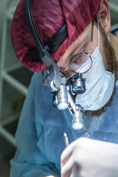
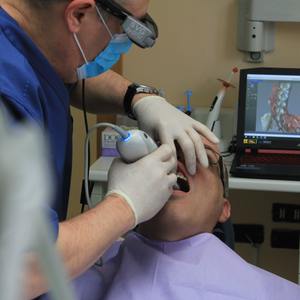
Implant placement is becoming more popular. Researchers at the University of Texas School of Dentistry at Houston discovered that careful management with cone-beam computed radiography is crucial for achieving instantaneous location. This is especially true in the lateral incisor region due to restricted access to alveolar bone.
The investigators agree that implant management can only be realized if there are facts and figures about the alveolar bone surrounding the site. While orthodox radiographic techniques were the standard for treatment preparation in the past, they are now questioned by imaging falsification or overlying.
Researchers found that the anterior maxilla's shape and general alveolar measurement had not been fully evaluated. The investigators found that the average alveolar measurement at anterior maxilla was 18.83 mm to 19.07mm in altitude, and 8.3mm to 9.62mm in thickness. The investigators also found that 41% of the central incisors and 77% of the lateral incisors have buccal undercuts of various distances. These results were comparable to information previously available for the mandibular posterior area, except for the high prevalence of undercuts in the lateral incisors. Because of the effect that alveolar shape and measurement will have on the implant placement and aesthetic results, the maxillary frontal region is the area that requires the most evaluation before operation. An insufficient transversal thickness of the crest would result in a decrease in span or render inclusion of implants impossible.


Comments (0)
See all